Recognizing Accredited Orthodontic Programs

Recognizing Accredited Orthodontic Programs

* Type of orthodontic treatment needed for the child.

Okay, so you're thinking about getting braces for your kid, right? Or maybe your teenager needs some orthodontic work. Either way, you want the best possible care, and that's totally understandable. That's where accreditation comes in, and honestly, it really matters, especially when we're talking about pediatric orthodontics.


Think of it like this: you wouldn't let just anyone perform surgery on your child, would you? You'd want a surgeon who's been properly trained and certified. Early orthodontic evaluations are recommended around age seven Youth orthodontic correction child. Accreditation in orthodontics is kind of similar. It's a stamp of approval that says an orthodontic program meets a very high standard of quality.


Recognizing accredited orthodontic programs means you're choosing a specialist who has not only graduated from dental school but has also completed a rigorous, two-to-three-year residency program specifically in orthodontics. These programs are evaluated by experts to make sure they're teaching the latest techniques, using the best equipment, and providing a comprehensive educational experience.


Why is that so important for kids? Well, children's mouths are still developing. An orthodontist who's gone through an accredited program is specifically trained to understand that growth and development, and they know how to gently guide teeth and jaws into the right positions. They're equipped to handle the unique challenges that come with treating young patients, like early intervention, growth modification, and even just making kids feel comfortable and safe in the dental chair.


Choosing an accredited program basically gives you peace of mind. You know your child is being treated by a highly qualified specialist who's committed to providing the best possible care. It's an investment in their smile, their confidence, and their overall oral health. And honestly, that's worth everything. So, do your homework, look for that accreditation, and make sure you're setting your child up for a lifetime of healthy, happy smiles.

So, you're trying to figure out if an orthodontic program is the real deal, right? Well, accreditation is your golden ticket. It's basically a stamp of approval saying the program meets really high standards. But what exactly makes an accredited program, well, accredited? It's not just about fancy chairs and gleaming instruments, though those are nice perks!


First off, you've gotta look at the faculty. Are they experienced orthodontists? Are they actively involved in research and the orthodontic community? Accredited programs usually boast a stellar faculty who are passionate about teaching and pushing the boundaries of the field. They're not just textbook regurgitators; they're mentors and leaders.


Then there's the curriculum. It needs to be comprehensive. We're talking a deep dive into everything from craniofacial growth and development to biomechanics and treatment planning. It's not just about straightening teeth; it's about understanding the whole system and how to create a stable, functional, and aesthetically pleasing result. The program has to cover a wide range of treatment modalities, too – braces, aligners, surgical orthodontics, interceptive treatment...the whole shebang.


Patient diversity is another huge piece of the puzzle. You want to be in a program where you're exposed to a wide range of cases, from simple malocclusions to complex craniofacial anomalies. This real-world experience is invaluable. You learn to adapt your skills and knowledge to different patient needs and challenges.


And finally, there's the emphasis on research and evidence-based practice. Orthodontics is constantly evolving, so a good program will encourage you to critically evaluate the literature, conduct your own research, and apply the latest scientific findings to your clinical practice. It's about being a lifelong learner and contributing to the advancement of the field.


Basically, an accredited orthodontic program isn't just about getting a certificate; it's about getting the best possible education and training to become a competent, ethical, and well-rounded orthodontist. It's an investment in your future and your patients' smiles. So, do your homework and choose wisely!

* Duration of the orthodontic treatment plan.

Finding the right orthodontist for your child is a big deal, and accreditation is a key factor. It's like making sure your child's school is up to par; you want the best, right? So, where do you even start looking for these orthodontists with the 'seal of approval,' so to speak?


Well, thankfully, there are resources out there to help navigate this process. The American Association of Orthodontists (AAO) is a fantastic place to begin. Their website often has a 'Find an Orthodontist' tool, allowing you to search specifically for AAO members in your area. AAO membership often, though not always, implies a commitment to maintaining the highest standards of care.


Another avenue to explore is asking your general dentist for recommendations. They often work closely with orthodontists and can provide valuable insights and suggest names of orthodontists they trust. Think of it as getting a referral from a trusted colleague.


Beyond these direct sources, online directories and review sites can be helpful, but approach them with a healthy dose of skepticism. Look for patterns in the reviews, rather than focusing on a single glowing or scathing comment. See if the orthodontist is mentioned in professional publications or has presented at conferences – these can be good indicators of their commitment to the field.


Ultimately, finding the right accredited orthodontist is a blend of research and personal connection. It's not just about the credentials on the wall; it's about finding someone who makes you and your child feel comfortable and confident in their care. So, use these resources as a starting point, but don't be afraid to schedule consultations and ask questions until you find the perfect fit.

* Geographic location and its cost of living.

Okay, so you're thinking about getting your teeth straightened, which is awesome! But before you hop into a chair and let someone start moving your pearly whites around, you gotta do your homework. And a big part of that homework is figuring out if the orthodontist you're considering actually knows their stuff. I mean, we're talking about your smile, your confidence, maybe even your long-term dental health. You don't want to leave that in the hands of someone who just dabbles in orthodontics on the weekends.


That's where accredited orthodontic programs come in. Think of it like this: it's the gold standard. It means the orthodontist has not just any dental degree, but has gone through extra, specialized training in straightening teeth and fixing bites. We're talking years of dedicated learning, hands-on experience, and a deep understanding of how teeth and jaws work together.


So, how do you find out if your potential orthodontist went through one of these top-notch programs? Easy, just ask! Don't be shy. It's your mouth, your money, and your smile on the line. Some questions you could ask are: "Where did you complete your orthodontic residency?" or "Is your program accredited by the American Dental Association's Commission on Dental Accreditation (CODA)?" If they hem and haw or can't give you a straight answer, that's a big red flag. You want someone who's proud of their training and happy to share the details.


Look, getting your teeth straightened is a big decision. Making sure your orthodontist has the right training is crucial. Accredited programs are there to give you peace of mind, knowing you're in good hands. So, ask those questions, do your research, and get ready to flash that confident, straight smile!

* Orthodontist's experience and specialization.

Okay, so you're thinking about straightening those pearly whites, huh? Maybe you've got a bit of a crooked grin, or perhaps your dentist mentioned a crossbite. Whatever the reason, you're starting to explore orthodontics. And that's smart! But here's the thing: not all orthodontists are created equal. That's where accreditation comes in.


Think of it like this: you wouldn't want someone tinkering under the hood of your car who just watched a YouTube video, right? You'd want a certified mechanic. Same deal with your teeth. An accredited orthodontist has gone through a rigorous program, a specialized residency after dental school, that focuses exclusively on moving teeth and correcting jaw alignment. They've seen it all, studied it all, and are equipped with the latest knowledge and techniques.


Why does this matter to you? Well, for starters, proper orthodontic treatment isn't just about aesthetics. Sure, a beautiful smile is a fantastic bonus, but it's also about improving your bite, making it easier to chew, speak clearly, and even prevent future problems like jaw pain and tooth wear. An accredited specialist understands all these nuances and can create a treatment plan that's tailored to your specific needs, not just a one-size-fits-all approach.


Choosing an accredited orthodontist also gives you peace of mind. You know they've met certain standards, that their education and training have been verified. You're putting your trust in a professional who's committed to providing the best possible care. It's an investment in your long-term oral health and well-being. So, do your homework, look for that accreditation, and get ready to flash a confident, healthy smile for years to come. It's worth it!

* Use of advanced technology or techniques.

Okay, let's talk about red flags when you're choosing an orthodontist. It's more than just straight teeth; it's about making sure you're in qualified hands. You're trusting someone with your smile, and that's a big deal. So, how do you tell if an orthodontist might be cutting corners or lacking the proper training?


First, be wary if they downplay the importance of board certification. A board-certified orthodontist has gone above and beyond, completing extra education and rigorous testing. It's not a mandatory requirement to practice, but it shows a real commitment to excellence. If an orthodontist seems dismissive of the American Board of Orthodontics (ABO) certification, or tries to make it seem unimportant, that should raise an eyebrow. It's not the only factor, but it's definitely a good sign of dedication and expertise.


Next, listen carefully to their treatment recommendations. A good orthodontist will thoroughly explain your options, the pros and cons of each, and why they recommend a particular approach. If they're pushing a specific treatment without explaining the alternatives, or if they seem vague or evasive about the process, be cautious. Transparency is key. You should feel like you understand what's happening and why.


Also, pay attention to the technology and techniques they use. Orthodontics is constantly evolving. While older methods can still be effective, a modern, well-accredited practice will usually incorporate newer technologies and techniques that can improve efficiency and comfort. If their office seems stuck in the past, or they're only offering very dated treatment options, it might suggest they haven't kept up with advancements in the field. Don't be afraid to ask about their continuing education and what new things they've learned recently.


Finally, trust your gut. If something feels off, it probably is. Maybe the office seems disorganized, the staff is unfriendly, or you just get a bad vibe. Choosing an orthodontist is a personal decision, and you should feel comfortable and confident in your choice. Don't hesitate to get a second opinion if you have any doubts. Your smile is worth it!

* Insurance coverage and payment options.

Okay, so you're thinking about braces for your kid, and you're probably swimming in information. Let's talk about something that might seem a bit dry, but it's actually super important: accreditation. Specifically, choosing an orthodontic program that's accredited.


Think of it this way: accreditation is like a gold star for orthodontic programs. It means a third-party organization, like the Commission on Dental Accreditation (CODA), has thoroughly checked out the program. They've made sure the curriculum is top-notch, the faculty are qualified, and that the students are getting the kind of training they need to become excellent orthodontists.


Now, why does this matter in the long run? Well, consider this: these future orthodontists are learning the latest techniques and technologies. They're being trained in a structured, rigorous environment that pushes them to be the best they can be. And where do they go after graduation? They go out into the world and start treating patients, often right in your own community.


Choosing an orthodontist who graduated from an accredited program gives you a little extra peace of mind. You know they've had solid training and are up-to-date on the best practices. It doesn't guarantee perfection – nobody can – but it significantly increases the odds of a successful outcome for your child's orthodontic treatment.


Beyond the technical aspects, accredited programs often emphasize patient care and ethical considerations. These orthodontists are not just learning how to straighten teeth; they're learning how to treat patients with respect and understanding. That's a big deal, especially when you're dealing with something as personal as your child's smile.


So, when you're researching orthodontists, take a little time to check where they received their training. Seeing that they graduated from an accredited program is a small detail that can make a big difference in the long run, contributing to a healthier, happier smile for your child. It's an investment in quality and a vote of confidence in the future of orthodontic care.

Crossbite
Unilateral posterior crossbite
Specialty Orthodontics

In dentistry, crossbite is a form of malocclusion where a tooth (or teeth) has a more buccal or lingual position (that is, the tooth is either closer to the cheek or to the tongue) than its corresponding antagonist tooth in the upper or lower dental arch. In other words, crossbite is a lateral misalignment of the dental arches.[1][2]

Anterior crossbite

[edit]
Class 1 with anterior crossbite

An anterior crossbite can be referred as negative overjet, and is typical of class III skeletal relations (prognathism).

Primary/mixed dentitions

[edit]

An anterior crossbite in a child with baby teeth or mixed dentition may happen due to either dental misalignment or skeletal misalignment. Dental causes may be due to displacement of one or two teeth, where skeletal causes involve either mandibular hyperplasia, maxillary hypoplasia or combination of both.

Dental crossbite

[edit]

An anterior crossbite due to dental component involves displacement of either maxillary central or lateral incisors lingual to their original erupting positions. This may happen due to delayed eruption of the primary teeth leading to permanent teeth moving lingual to their primary predecessors. This will lead to anterior crossbite where upon biting, upper teeth are behind the lower front teeth and may involve few or all frontal incisors. In this type of crossbite, the maxillary and mandibular proportions are normal to each other and to the cranial base. Another reason that may lead to a dental crossbite is crowding in the maxillary arch. Permanent teeth will tend to erupt lingual to the primary teeth in presence of crowding. Side-effects caused by dental crossbite can be increased recession on the buccal of lower incisors and higher chance of inflammation in the same area. Another term for an anterior crossbite due to dental interferences is Pseudo Class III Crossbite or Malocclusion.

Single tooth crossbite

[edit]

Single tooth crossbites can occur due to uneruption of a primary teeth in a timely manner which causes permanent tooth to erupt in a different eruption pattern which is lingual to the primary tooth.[3] Single tooth crossbites are often fixed by using a finger-spring based appliances.[4][5] This type of spring can be attached to a removable appliance which is used by patient every day to correct the tooth position.

Skeletal crossbite

[edit]

An anterior crossbite due to skeletal reasons will involve a deficient maxilla and a more hyperplastic or overgrown mandible. People with this type of crossbite will have dental compensation which involves proclined maxillary incisors and retroclined mandibular incisors. A proper diagnosis can be made by having a person bite into their centric relation will show mandibular incisors ahead of the maxillary incisors, which will show the skeletal discrepancy between the two jaws.[6]

Posterior crossbite

[edit]

Bjork defined posterior crossbite as a malocclusion where the buccal cusps of canine, premolar and molar of upper teeth occlude lingually to the buccal cusps of canine, premolar and molar of lower teeth.[7] Posterior crossbite is often correlated to a narrow maxilla and upper dental arch. A posterior crossbite can be unilateral, bilateral, single-tooth or entire segment crossbite. Posterior crossbite has been reported to occur between 7–23% of the population.[8][9] The most common type of posterior crossbite to occur is the unilateral crossbite which occurs in 80% to 97% of the posterior crossbite cases.[10][3] Posterior crossbites also occur most commonly in primary and mixed dentition. This type of crossbite usually presents with a functional shift of the mandible towards the side of the crossbite. Posterior crossbite can occur due to either skeletal, dental or functional abnormalities. One of the common reasons for development of posterior crossbite is the size difference between maxilla and mandible, where maxilla is smaller than mandible.[11] Posterior crossbite can result due to

  • Upper Airway Obstruction where people with "adenoid faces" who have trouble breathing through their nose. They have an open bite malocclusion and present with development of posterior crossbite.[12]
  • Prolong digit or suckling habits which can lead to constriction of maxilla posteriorly[13]
  • Prolong pacifier use (beyond age 4)[13]

Connections with TMD

[edit]

Unilateral posterior crossbite

[edit]

Unilateral crossbite involves one side of the arch. The most common cause of unilateral crossbite is a narrow maxillary dental arch. This can happen due to habits such as digit sucking, prolonged use of pacifier or upper airway obstruction. Due to the discrepancy between the maxillary and mandibular arch, neuromuscular guidance of the mandible causes mandible to shift towards the side of the crossbite.[14] This is also known as Functional mandibular shift. This shift can become structural if left untreated for a long time during growth, leading to skeletal asymmetries. Unilateral crossbites can present with following features in a child

  • Lower midline deviation[15] to the crossbite side
  • Class 2 Subdivision relationships
  • Temporomandibular disorders [16]

Treatment

[edit]

A child with posterior crossbite should be treated immediately if the child shifts their mandible on closing, which is often seen in a unilateral crossbite as mentioned above. The best age to treat a child with crossbite is in their mixed dentition when their palatal sutures have not fused to each other. Palatal expansion allows more space in an arch to relieve crowding and correct posterior crossbite. The correction can include any type of palatal expanders that will expand the palate which resolves the narrow constriction of the maxilla.[9] There are several therapies that can be used to correct a posterior crossbite: braces, 'Z' spring or cantilever spring, quad helix, removable plates, clear aligner therapy, or a Delaire mask. The correct therapy should be decided by the orthodontist depending on the type and severity of the crossbite.

One of the keys in diagnosing the anterior crossbite due to skeletal vs dental causes is diagnosing a CR-CO shift in a patient. An adolescent presenting with anterior crossbite may be positioning their mandible forward into centric occlusion (CO) due to the dental interferences. Thus finding their occlusion in centric relation (CR) is key in diagnosis. For anterior crossbite, if their CO matches their CR then the patient truly has a skeletal component to their crossbite. If the CR shows a less severe class 3 malocclusion or teeth not in anterior crossbite, this may mean that their anterior crossbite results due to dental interferences.[17]

Goal to treat unilateral crossbites should definitely include removal of occlusal interferences and elimination of the functional shift. Treating posterior crossbites early may help prevent the occurrence of Temporomandibular joint pathology.[18]

Unilateral crossbites can also be diagnosed and treated properly by using a Deprogramming splint. This splint has flat occlusal surface which causes the muscles to deprogram themselves and establish new sensory engrams. When the splint is removed, a proper centric relation bite can be diagnosed from the bite.[19]

Self-correction

[edit]

Literature states that very few crossbites tend to self-correct which often justify the treatment approach of correcting these bites as early as possible.[9] Only 0–9% of crossbites self-correct. Lindner et al. reported that 50% of crossbites were corrected in 76 four-year-old children.[20]

See also

[edit]
  • List of palatal expanders
  • Palatal expansion
  • Malocclusion

References

[edit]
  1. ^ "Elsevier: Proffit: Contemporary Orthodontics · Welcome". www.contemporaryorthodontics.com. Retrieved 2016-12-11.
  2. ^ Borzabadi-Farahani A, Borzabadi-Farahani A, Eslamipour F (October 2009). "Malocclusion and occlusal traits in an urban Iranian population. An epidemiological study of 11- to 14-year-old children". European Journal of Orthodontics. 31 (5): 477–84. doi:10.1093/ejo/cjp031. PMID 19477970.
  3. ^ a b Kutin, George; Hawes, Roland R. (1969-11-01). "Posterior cross-bites in the deciduous and mixed dentitions". American Journal of Orthodontics. 56 (5): 491–504. doi:10.1016/0002-9416(69)90210-3. PMID 5261162.
  4. ^ Zietsman, S. T.; Visagé, W.; Coetzee, W. J. (2000-11-01). "Palatal finger springs in removable orthodontic appliances--an in vitro study". South African Dental Journal. 55 (11): 621–627. ISSN 1029-4864. PMID 12608226.
  5. ^ Ulusoy, Ayca Tuba; Bodrumlu, Ebru Hazar (2013-01-01). "Management of anterior dental crossbite with removable appliances". Contemporary Clinical Dentistry. 4 (2): 223–226. doi:10.4103/0976-237X.114855. ISSN 0976-237X. PMC 3757887. PMID 24015014.
  6. ^ Al-Hummayani, Fadia M. (2017-03-05). "Pseudo Class III malocclusion". Saudi Medical Journal. 37 (4): 450–456. doi:10.15537/smj.2016.4.13685. ISSN 0379-5284. PMC 4852025. PMID 27052290.
  7. ^ Bjoerk, A.; Krebs, A.; Solow, B. (1964-02-01). "A Method for Epidemiological Registration of Malocculusion". Acta Odontologica Scandinavica. 22: 27–41. doi:10.3109/00016356408993963. ISSN 0001-6357. PMID 14158468.
  8. ^ Moyers, Robert E. (1988-01-01). Handbook of orthodontics. Year Book Medical Publishers. ISBN 9780815160038.
  9. ^ a b c Thilander, Birgit; Lennartsson, Bertil (2002-09-01). "A study of children with unilateral posterior crossbite, treated and untreated, in the deciduous dentition--occlusal and skeletal characteristics of significance in predicting the long-term outcome". Journal of Orofacial Orthopedics. 63 (5): 371–383. doi:10.1007/s00056-002-0210-6. ISSN 1434-5293. PMID 12297966. S2CID 21857769.
  10. ^ Thilander, Birgit; Wahlund, Sonja; Lennartsson, Bertil (1984-01-01). "The effect of early interceptive treatment in children with posterior cross-bite". The European Journal of Orthodontics. 6 (1): 25–34. doi:10.1093/ejo/6.1.25. ISSN 0141-5387. PMID 6583062.
  11. ^ Allen, David; Rebellato, Joe; Sheats, Rose; Ceron, Ana M. (2003-10-01). "Skeletal and dental contributions to posterior crossbites". The Angle Orthodontist. 73 (5): 515–524. ISSN 0003-3219. PMID 14580018.
  12. ^ Bresolin, D.; Shapiro, P. A.; Shapiro, G. G.; Chapko, M. K.; Dassel, S. (1983-04-01). "Mouth breathing in allergic children: its relationship to dentofacial development". American Journal of Orthodontics. 83 (4): 334–340. doi:10.1016/0002-9416(83)90229-4. ISSN 0002-9416. PMID 6573147.
  13. ^ a b Ogaard, B.; Larsson, E.; Lindsten, R. (1994-08-01). "The effect of sucking habits, cohort, sex, intercanine arch widths, and breast or bottle feeding on posterior crossbite in Norwegian and Swedish 3-year-old children". American Journal of Orthodontics and Dentofacial Orthopedics. 106 (2): 161–166. doi:10.1016/S0889-5406(94)70034-6. ISSN 0889-5406. PMID 8059752.
  14. ^ Piancino, Maria Grazia; Kyrkanides, Stephanos (2016-04-18). Understanding Masticatory Function in Unilateral Crossbites. John Wiley & Sons. ISBN 9781118971871.
  15. ^ Brin, Ilana; Ben-Bassat, Yocheved; Blustein, Yoel; Ehrlich, Jacob; Hochman, Nira; Marmary, Yitzhak; Yaffe, Avinoam (1996-02-01). "Skeletal and functional effects of treatment for unilateral posterior crossbite". American Journal of Orthodontics and Dentofacial Orthopedics. 109 (2): 173–179. doi:10.1016/S0889-5406(96)70178-6. PMID 8638566.
  16. ^ Pullinger, A. G.; Seligman, D. A.; Gornbein, J. A. (1993-06-01). "A multiple logistic regression analysis of the risk and relative odds of temporomandibular disorders as a function of common occlusal features". Journal of Dental Research. 72 (6): 968–979. doi:10.1177/00220345930720061301. ISSN 0022-0345. PMID 8496480. S2CID 25351006.
  17. ^ COSTEA, CARMEN MARIA; BADEA, MÎNDRA EUGENIA; VASILACHE, SORIN; MESAROÅž, MICHAELA (2016-01-01). "Effects of CO-CR discrepancy in daily orthodontic treatment planning". Clujul Medical. 89 (2): 279–286. doi:10.15386/cjmed-538. ISSN 1222-2119. PMC 4849388. PMID 27152081.
  18. ^ Kennedy, David B.; Osepchook, Matthew (2005-09-01). "Unilateral posterior crossbite with mandibular shift: a review". Journal (Canadian Dental Association). 71 (8): 569–573. ISSN 1488-2159. PMID 16202196.
  19. ^ Nielsen, H. J.; Bakke, M.; Blixencrone-Møller, T. (1991-12-01). "[Functional and orthodontic treatment of a patient with an open bite craniomandibular disorder]". Tandlaegebladet. 95 (18): 877–881. ISSN 0039-9353. PMID 1817382.
  20. ^ Lindner, A. (1989-10-01). "Longitudinal study on the effect of early interceptive treatment in 4-year-old children with unilateral cross-bite". Scandinavian Journal of Dental Research. 97 (5): 432–438. doi:10.1111/j.1600-0722.1989.tb01457.x. ISSN 0029-845X. PMID 2617141.
[edit]


Tooth
A chimpanzee displaying his teeth
Details
Identifiers
Latin dens
MeSH D014070
FMA 12516
Anatomical terminology
[edit on Wikidata]

A tooth (pl.: teeth) is a hard, calcified structure found in the jaws (or mouths) of many vertebrates and used to break down food. Some animals, particularly carnivores and omnivores, also use teeth to help with capturing or wounding prey, tearing food, for defensive purposes, to intimidate other animals often including their own, or to carry prey or their young. The roots of teeth are covered by gums. Teeth are not made of bone, but rather of multiple tissues of varying density and hardness that originate from the outermost embryonic germ layer, the ectoderm.

The general structure of teeth is similar across the vertebrates, although there is considerable variation in their form and position. The teeth of mammals have deep roots, and this pattern is also found in some fish, and in crocodilians. In most teleost fish, however, the teeth are attached to the outer surface of the bone, while in lizards they are attached to the inner surface of the jaw by one side. In cartilaginous fish, such as sharks, the teeth are attached by tough ligaments to the hoops of cartilage that form the jaw.[1]

Monophyodonts are animals that develop only one set of teeth, while diphyodonts grow an early set of deciduous teeth and a later set of permanent or "adult" teeth. Polyphyodonts grow many sets of teeth. For example, sharks, grow a new set of teeth every two weeks to replace worn teeth. Most extant mammals including humans are diphyodonts, but there are exceptions including elephants, kangaroos, and manatees, all of which are polyphyodonts.

Rodent incisors grow and wear away continually through gnawing, which helps maintain relatively constant length. The industry of the beaver is due in part to this qualification. Some rodents, such as voles and guinea pigs (but not mice), as well as lagomorpha (rabbits, hares and pikas), have continuously growing molars in addition to incisors.[2][3] Also, tusks (in tusked mammals) grow almost throughout life.[4]

Teeth are not always attached to the jaw, as they are in mammals. In many reptiles and fish, teeth are attached to the palate or to the floor of the mouth, forming additional rows inside those on the jaws proper. Some teleosts even have teeth in the pharynx. While not true teeth in the usual sense, the dermal denticles of sharks are almost identical in structure and are likely to have the same evolutionary origin. Indeed, teeth appear to have first evolved in sharks, and are not found in the more primitive jawless fish – while lampreys do have tooth-like structures on the tongue, these are in fact, composed of keratin, not of dentine or enamel, and bear no relationship to true teeth.[1] Though "modern" teeth-like structures with dentine and enamel have been found in late conodonts, they are now supposed to have evolved independently of later vertebrates' teeth.[5][6]

Living amphibians typically have small teeth, or none at all, since they commonly feed only on soft foods. In reptiles, teeth are generally simple and conical in shape, although there is some variation between species, most notably the venom-injecting fangs of snakes. The pattern of incisors, canines, premolars and molars is found only in mammals, and to varying extents, in their evolutionary ancestors. The numbers of these types of teeth vary greatly between species; zoologists use a standardised dental formula to describe the precise pattern in any given group.[1]

Etymology

[edit]

The word tooth comes from Proto-Germanic *tanþs, derived from the Proto-Indo-European *h₁dent-, which was composed of the root *h₁ed- 'to eat' plus the active participle suffix *-nt, therefore literally meaning 'that which eats'.[7]

The irregular plural form teeth is the result of Germanic umlaut whereby vowels immediately preceding a high vocalic in the following syllable were raised. As the nominative plural ending of the Proto-Germanic consonant stems (to which *tanþs belonged) was *-iz, the root vowel in the plural form *tanþiz (changed by this point to *tÄ…Ì„þi via unrelated phonological processes) was raised to /œÃƒÆ’ƒÆ’‹Â/, and later unrounded to /eː/, resulting in the tōþ/tÄ“þ alternation attested from Old English. Cf. also Old English bōc/bēċ 'book/books' and 'mÅ«s/mȳs' 'mouse/mice', from Proto-Germanic *bōks/bōkiz and *mÅ«s/mÅ«siz respectively.

Cognate with Latin dÄ“ns, Greek á½€δούς (odous), and Sanskrit dát.

Origin

[edit]

Teeth are assumed to have evolved either from ectoderm denticles (scales, much like those on the skin of sharks) that folded and integrated into the mouth (called the "outside–in" theory), or from endoderm pharyngeal teeth (primarily formed in the pharynx of jawless vertebrates) (the "inside–out" theory). In addition, there is another theory stating that neural crest gene regulatory network, and neural crest-derived ectomesenchyme are the key to generate teeth (with any epithelium, either ectoderm or endoderm).[4][8]

The genes governing tooth development in mammals are homologous to those involved in the development of fish scales.[9] Study of a tooth plate of a fossil of the extinct fish Romundina stellina showed that the teeth and scales were made of the same tissues, also found in mammal teeth, lending support to the theory that teeth evolved as a modification of scales.[10]

Mammals

[edit]

Teeth are among the most distinctive (and long-lasting) features of mammal species. Paleontologists use teeth to identify fossil species and determine their relationships. The shape of the animal's teeth are related to its diet. For example, plant matter is hard to digest, so herbivores have many molars for chewing and grinding. Carnivores, on the other hand, have canine teeth to kill prey and to tear meat.

Mammals, in general, are diphyodont, meaning that they develop two sets of teeth. In humans, the first set (the "baby", "milk", "primary" or "deciduous" set) normally starts to appear at about six months of age, although some babies are born with one or more visible teeth, known as neonatal teeth. Normal tooth eruption at about six months is known as teething and can be painful. Kangaroos, elephants, and manatees are unusual among mammals because they are polyphyodonts.

Aardvark

[edit]

In aardvarks, teeth lack enamel and have many pulp tubules, hence the name of the order Tubulidentata.[11]

Canines

[edit]

In dogs, the teeth are less likely than humans to form dental cavities because of the very high pH of dog saliva, which prevents enamel from demineralizing.[12] Sometimes called cuspids, these teeth are shaped like points (cusps) and are used for tearing and grasping food.[13]

Cetaceans

[edit]

Like human teeth, whale teeth have polyp-like protrusions located on the root surface of the tooth. These polyps are made of cementum in both species, but in human teeth, the protrusions are located on the outside of the root, while in whales the nodule is located on the inside of the pulp chamber. While the roots of human teeth are made of cementum on the outer surface, whales have cementum on the entire surface of the tooth with a very small layer of enamel at the tip. This small enamel layer is only seen in older whales where the cementum has been worn away to show the underlying enamel.[14]

The toothed whale is a parvorder of the cetaceans characterized by having teeth. The teeth differ considerably among the species. They may be numerous, with some dolphins bearing over 100 teeth in their jaws. On the other hand, the narwhals have a giant unicorn-like tusk, which is a tooth containing millions of sensory pathways and used for sensing during feeding, navigation, and mating. It is the most neurologically complex tooth known. Beaked whales are almost toothless, with only bizarre teeth found in males. These teeth may be used for feeding but also for demonstrating aggression and showmanship.

Primates

[edit]

In humans (and most other primates), there are usually 20 primary (also "baby" or "milk") teeth, and later up to 32 permanent teeth. Four of these 32 may be third molars or wisdom teeth, although these are not present in all adults, and may be removed surgically later in life.[15]

Among primary teeth, 10 of them are usually found in the maxilla (i.e. upper jaw) and the other 10 in the mandible (i.e. lower jaw). Among permanent teeth, 16 are found in the maxilla and the other 16 in the mandible. Most of the teeth have uniquely distinguishing features.

Horse

[edit]

An adult horse has between 36 and 44 teeth. The enamel and dentin layers of horse teeth are intertwined.[16] All horses have 12 premolars, 12 molars, and 12 incisors.[17] Generally, all male equines also have four canine teeth (called tushes) between the molars and incisors. However, few female horses (less than 28%) have canines, and those that do usually have only one or two, which many times are only partially erupted.[18] A few horses have one to four wolf teeth, which are vestigial premolars, with most of those having only one or two. They are equally common in male and female horses and much more likely to be on the upper jaw. If present these can cause problems as they can interfere with the horse's bit contact. Therefore, wolf teeth are commonly removed.[17]

Horse teeth can be used to estimate the animal's age. Between birth and five years, age can be closely estimated by observing the eruption pattern on milk teeth and then permanent teeth. By age five, all permanent teeth have usually erupted. The horse is then said to have a "full" mouth. After the age of five, age can only be conjectured by studying the wear patterns on the incisors, shape, the angle at which the incisors meet, and other factors. The wear of teeth may also be affected by diet, natural abnormalities, and cribbing. Two horses of the same age may have different wear patterns.

A horse's incisors, premolars, and molars, once fully developed, continue to erupt as the grinding surface is worn down through chewing. A young adult horse will have teeth, which are 110–130 mm (4.5–5 inches) long, with the majority of the crown remaining below the gumline in the dental socket. The rest of the tooth will slowly emerge from the jaw, erupting about 3 mm (18 in) each year, as the horse ages. When the animal reaches old age, the crowns of the teeth are very short and the teeth are often lost altogether. Very old horses, if lacking molars, may need to have their fodder ground up and soaked in water to create a soft mush for them to eat in order to obtain adequate nutrition.

Proboscideans

[edit]
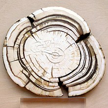
Section through the ivory tusk of a mammoth

Elephants' tusks are specialized incisors for digging food up and fighting. Some elephant teeth are similar to those in manatees, and elephants are believed to have undergone an aquatic phase in their evolution.

At birth, elephants have a total of 28 molar plate-like grinding teeth not including the tusks. These are organized into four sets of seven successively larger teeth which the elephant will slowly wear through during its lifetime of chewing rough plant material. Only four teeth are used for chewing at a given time, and as each tooth wears out, another tooth moves forward to take its place in a process similar to a conveyor belt. The last and largest of these teeth usually becomes exposed when the animal is around 40 years of age, and will often last for an additional 20 years. When the last of these teeth has fallen out, regardless of the elephant's age, the animal will no longer be able to chew food and will die of starvation.[19][20]

Rabbit

[edit]

Rabbits and other lagomorphs usually shed their deciduous teeth before (or very shortly after) their birth, and are usually born with their permanent teeth.[21] The teeth of rabbits complement their diet, which consists of a wide range of vegetation. Since many of the foods are abrasive enough to cause attrition, rabbit teeth grow continuously throughout life.[22] Rabbits have a total of six incisors, three upper premolars, three upper molars, two lower premolars, and two lower molars on each side. There are no canines. Dental formula is 2.0.3.31.0.2.3 = 28. Three to four millimeters of the tooth is worn away by incisors every week, whereas the cheek teeth require a month to wear away the same amount.[23]

The incisors and cheek teeth of rabbits are called aradicular hypsodont teeth. This is sometimes referred to as an elodent dentition. These teeth grow or erupt continuously. The growth or eruption is held in balance by dental abrasion from chewing a diet high in fiber.

Buccal view of top incisor from Rattus rattus. Top incisor outlined in yellow. Molars circled in blue.
Buccal view of the lower incisor from the right dentary of a Rattus rattus
Lingual view of the lower incisor from the right dentary of a Rattus rattus
Midsagittal view of top incisor from Rattus rattus. Top incisor outlined in yellow. Molars circled in blue.

Rodents

[edit]

Rodents have upper and lower hypselodont incisors that can continuously grow enamel throughout its life without having properly formed roots.[24] These teeth are also known as aradicular teeth, and unlike humans whose ameloblasts die after tooth development, rodents continually produce enamel, they must wear down their teeth by gnawing on various materials.[25] Enamel and dentin are produced by the enamel organ, and growth is dependent on the presence of stem cells, cellular amplification, and cellular maturation structures in the odontogenic region.[26] Rodent incisors are used for cutting wood, biting through the skin of fruit, or for defense. This allows for the rate of wear and tooth growth to be at equilibrium.[24] The microstructure of rodent incisor enamel has shown to be useful in studying the phylogeny and systematics of rodents because of its independent evolution from the other dental traits. The enamel on rodent incisors are composed of two layers: the inner portio interna (PI) with Hunter-Schreger bands (HSB) and an outer portio externa (PE) with radial enamel (RE).[27] It usually involves the differential regulation of the epithelial stem cell niche in the tooth of two rodent species, such as guinea pigs.[28][29]

Lingual view of top incisor from Rattus rattus. Top incisor outlined in yellow. Molars circled in blue.

The teeth have enamel on the outside and exposed dentin on the inside, so they self-sharpen during gnawing. On the other hand, continually growing molars are found in some rodent species, such as the sibling vole and the guinea pig.[28][29] There is variation in the dentition of the rodents, but generally, rodents lack canines and premolars, and have a space between their incisors and molars, called the diastema region.

Manatee

[edit]

Manatees are polyphyodont with mandibular molars developing separately from the jaw and are encased in a bony shell separated by soft tissue.[30][31]

Walrus

[edit]

Walrus tusks are canine teeth that grow continuously throughout life.[32]

Fish

[edit]
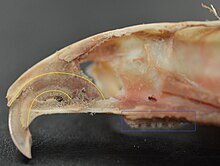
Teeth of a great white shark

Fish, such as sharks, may go through many teeth in their lifetime. The replacement of multiple teeth is known as polyphyodontia.

A class of prehistoric shark are called cladodonts for their strange forked teeth.

Unlike the continuous shedding of functional teeth seen in modern sharks,[33][34] the majority of stem chondrichthyan lineages retained all tooth generations developed throughout the life of the animal.[35] This replacement mechanism is exemplified by the tooth whorl-based dentitions of acanthodians,[36] which include the oldest known toothed vertebrate, Qianodus duplicis[37].

Amphibians

[edit]

All amphibians have pedicellate teeth, which are modified to be flexible due to connective tissue and uncalcified dentine that separates the crown from the base of the tooth.[38]

Most amphibians exhibit teeth that have a slight attachment to the jaw or acrodont teeth. Acrodont teeth exhibit limited connection to the dentary and have little enervation.[39] This is ideal for organisms who mostly use their teeth for grasping, but not for crushing and allows for rapid regeneration of teeth at a low energy cost. Teeth are usually lost in the course of feeding if the prey is struggling. Additionally, amphibians that undergo a metamorphosis develop bicuspid shaped teeth.[40]

Reptiles

[edit]

The teeth of reptiles are replaced constantly throughout their lives. Crocodilian juveniles replace teeth with larger ones at a rate as high as one new tooth per socket every month. Once mature, tooth replacement rates can slow to two years and even longer. Overall, crocodilians may use 3,000 teeth from birth to death. New teeth are created within old teeth.[41]

Birds

[edit]

A skull of Ichthyornis discovered in 2014 suggests that the beak of birds may have evolved from teeth to allow chicks to escape their shells earlier, and thus avoid predators and also to penetrate protective covers such as hard earth to access underlying food.[42][43]

Invertebrates

[edit]
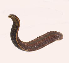
The European medicinal leech has three jaws with numerous sharp teeth which function like little saws for incising a host.

True teeth are unique to vertebrates,[44] although many invertebrates have analogous structures often referred to as teeth. The organisms with the simplest genome bearing such tooth-like structures are perhaps the parasitic worms of the family Ancylostomatidae.[45] For example, the hookworm Necator americanus has two dorsal and two ventral cutting plates or teeth around the anterior margin of the buccal capsule. It also has a pair of subdorsal and a pair of subventral teeth located close to the rear.[46]

Historically, the European medicinal leech, another invertebrate parasite, has been used in medicine to remove blood from patients.[47] They have three jaws (tripartite) that resemble saws in both appearance and function, and on them are about 100 sharp teeth used to incise the host. The incision leaves a mark that is an inverted Y inside of a circle. After piercing the skin and injecting anticoagulants (hirudin) and anaesthetics, they suck out blood, consuming up to ten times their body weight in a single meal.[48]

In some species of Bryozoa, the first part of the stomach forms a muscular gizzard lined with chitinous teeth that crush armoured prey such as diatoms. Wave-like peristaltic contractions then move the food through the stomach for digestion.[49]

The limpet rasps algae from rocks using teeth with the strongest known tensile strength of any biological material.

Molluscs have a structure called a radula, which bears a ribbon of chitinous teeth. However, these teeth are histologically and developmentally different from vertebrate teeth and are unlikely to be homologous. For example, vertebrate teeth develop from a neural crest mesenchyme-derived dental papilla, and the neural crest is specific to vertebrates, as are tissues such as enamel.[44]

The radula is used by molluscs for feeding and is sometimes compared rather inaccurately to a tongue. It is a minutely toothed, chitinous ribbon, typically used for scraping or cutting food before the food enters the oesophagus. The radula is unique to molluscs, and is found in every class of mollusc apart from bivalves.

Within the gastropods, the radula is used in feeding by both herbivorous and carnivorous snails and slugs. The arrangement of teeth (also known as denticles) on the radula ribbon varies considerably from one group to another as shown in the diagram on the left.

Predatory marine snails such as the Naticidae use the radula plus an acidic secretion to bore through the shell of other molluscs. Other predatory marine snails, such as the Conidae, use a specialized radula tooth as a poisoned harpoon. Predatory pulmonate land slugs, such as the ghost slug, use elongated razor-sharp teeth on the radula to seize and devour earthworms. Predatory cephalopods, such as squid, use the radula for cutting prey.

In most of the more ancient lineages of gastropods, the radula is used to graze by scraping diatoms and other microscopic algae off rock surfaces and other substrates. Limpets scrape algae from rocks using radula equipped with exceptionally hard rasping teeth.[50] These teeth have the strongest known tensile strength of any biological material, outperforming spider silk.[50] The mineral protein of the limpet teeth can withstand a tensile stress of 4.9 GPa, compared to 4 GPa of spider silk and 0.5 GPa of human teeth.[51]


Fossilization and taphonomy

[edit]

Because teeth are very resistant, often preserved when bones are not,[52] and reflect the diet of the host organism, they are very valuable to archaeologists and palaeontologists.[53] Early fish such as the thelodonts had scales composed of dentine and an enamel-like compound, suggesting that the origin of teeth was from scales which were retained in the mouth. Fish as early as the late Cambrian had dentine in their exoskeletons, which may have functioned in defense or for sensing their environments.[54] Dentine can be as hard as the rest of teeth and is composed of collagen fibres, reinforced with hydroxyapatite.[54]

Though teeth are very resistant, they also can be brittle and highly susceptible to cracking.[55] However, cracking of the tooth can be used as a diagnostic tool for predicting bite force. Additionally, enamel fractures can also give valuable insight into the diet and behaviour of archaeological and fossil samples.

Decalcification removes the enamel from teeth and leaves only the organic interior intact, which comprises dentine and cementine.[56] Enamel is quickly decalcified in acids,[57] perhaps by dissolution by plant acids or via diagenetic solutions, or in the stomachs of vertebrate predators.[56] Enamel can be lost by abrasion or spalling,[56] and is lost before dentine or bone are destroyed by the fossilisation process.[57] In such a case, the 'skeleton' of the teeth would consist of the dentine, with a hollow pulp cavity.[56] The organic part of dentine, conversely, is destroyed by alkalis.[57]

See also

[edit]
  • Animal tooth development
  • Dragon's teeth (mythology)

References

[edit]
  1. ^ a b c Romer, Alfred Sherwood; Parsons, Thomas S. (1977). The Vertebrate Body. Philadelphia, PA: Holt-Saunders International. pp. 300–310. ISBN 978-0-03-910284-5.
  2. ^ Tummers M, Thesleff I (March 2003). "Root or crown: a developmental choice orchestrated by the differential regulation of the epithelial stem cell niche in the tooth of two rodent species". Development. 130 (6): 1049–57. doi:10.1242/dev.00332. PMID 12571097.
  3. ^ Hunt AM (1959). "A description of the molar teeth and investing tissues of normal guinea pigs". J. Dent. Res. 38 (2): 216–31. doi:10.1177/00220345590380020301. PMID 13641521. S2CID 45097018.
  4. ^ a b Nasoori, Alireza (2020). "Tusks, the extra-oral teeth". Archives of Oral Biology. 117: 104835. doi:10.1016/j.archoralbio.2020.104835. PMID 32668361. S2CID 220585014.
  5. ^ McCOLLUM, MELANIE; SHARPE, PAUL T. (July 2001). "Evolution and development of teeth". Journal of Anatomy. 199 (1–2): 153–159. doi:10.1046/j.1469-7580.2001.19910153.x. PMC 1594990. PMID 11523817.
  6. ^ Kaplan, Matt (October 16, 2013). "Fossil scans reveal origins of teeth". Nature. doi:10.1038/nature.2013.13964 – via www.nature.com.
  7. ^ Harper, Douglas (2001–2021). "tooth | Origin and meaning of tooth". Online Etymology Dictionary.
  8. ^ Jheon, Andrew H (2012). "From molecules to mastication: the development and evolution of teeth". Wiley Interdiscip Rev Dev Biol. 2 (2): 165–182. doi:10.1002/wdev.63. PMC 3632217. PMID 24009032.
  9. ^ Sharpe, P. T. (2001). "Fish scale development: Hair today, teeth and scales yesterday?". Current Biology. 11 (18): R751 – R752. Bibcode:2001CBio...11.R751S. doi:10.1016/S0960-9822(01)00438-9. PMID 11566120. S2CID 18868124.
  10. ^ Jennifer Viegas (June 24, 2015). "First-known teeth belonged to fierce fish". ABC Science. Retrieved June 28, 2015.
  11. ^ Shoshani 2002, p. 619
  12. ^ Hale, FA (2009). "Dental caries in the dog". Can. Vet. J. 50 (12): 1301–4. PMC 2777300. PMID 20190984.
  13. ^ "Types of Teeth, Dental Anatomy & Tooth Anatomy | Colgate®". www.colgate.com. Archived from the original on 2017-11-19. Retrieved 2017-11-19.
  14. ^ "Common Characteristics Of Whale Teeth". Archived from the original on 4 September 2011. Retrieved 18 July 2014.
  15. ^ "Everything you need to know about teeth". NHS Scotland. Retrieved 5 May 2020.
  16. ^ "Gummed Out: Young Horses Lose Many Teeth, Vet Says". Archived from the original on 8 July 2014. Retrieved 6 July 2014.
  17. ^ a b Patricia Pence (2002). Equine Dentistry: A Practical Guide. Baltimore: Lippincott Williams & Wilkins. ISBN 978-0-683-30403-9.
  18. ^ Al Cirelli. "Equine Dentition" (PDF). University of Nevada Reno. SP-00-08. Retrieved 7 June 2010.
  19. ^ Maurice Burton; Robert Burton (2002). International Wildlife Encyclopedia. Marshall Cavendish. p. 769. ISBN 978-0-7614-7266-7.
  20. ^ Bram, L. et al. MCMLXXXIII. Elephants. Funk & Wagnalls New Encyclopedia, Volume 9, p. 183. ISBN 0-8343-0051-6
  21. ^ "Dental Anatomy & Care for Rabbits and Rodents".
  22. ^ Brown, Susan. Rabbit Dental Diseases Archived 2007-10-14 at the Wayback Machine, hosted on the San Diego Chapter of the House Rabbit Society Archived 2007-10-13 at the Wayback Machine. Page accessed April 9, 2007.
  23. ^ Ryšavy, Robin. Hay & Dental Health, hosted by the Missouri House Rabbit Society-Kansas City. Page accessed January 2, 2024.
  24. ^ a b Cox, Philip; Hautier, Lionel (2015). Evolution of the Rodents: Advances in Phylogeny, Functional Morphology and Development. Cambridge University Press. p. 482. ISBN 9781107044333.
  25. ^ Caceci, Thomas. Veterinary Histology with subtitle "Digestive System: Oral Cavity" found here Archived 2006-04-30 at the Wayback Machine.
  26. ^ Gomes, J.r.; Omar, N.f.; Do Carmo, E.r.; Neves, J.s.; Soares, M.a.m.; Narvaes, E.a.; Novaes, P.d. (30 April 2013). "Relationship Between Cell Proliferation and Eruption Rate in the Rat Incisor". The Anatomical Record. 296 (7): 1096–1101. doi:10.1002/ar.22712. ISSN 1932-8494. PMID 23629828. S2CID 13197331.
  27. ^ Martin, Thomas (September 1999). "Evolution of Incisor Enamel Microstructure in Theridomyidae (Rodentia)". Journal of Vertebrate Paleontology. 19 (3): 550. Bibcode:1999JVPal..19..550M. doi:10.1080/02724634.1999.10011164.
  28. ^ a b Tummers M and Thesleff I. Root or crown: a developmental choice orchestrated by the differential regulation of the epithelial stem cell niche in the tooth of two rodent species. Development (2003). 130(6):1049-57.
  29. ^ a b AM Hunt. A description of the molar teeth and investing tissues of normal guinea pigs. J Dent Res. (1959) 38(2):216-31.
  30. ^ Shoshani, J., ed. (2000). Elephants: Majestic Creatures of the Wild. Checkmark Books. ISBN 0-87596-143-6.
  31. ^ Best, Robin (1984). Macdonald, D. (ed.). The Encyclopedia of Mammals. New York: Facts on File. pp. 292–298. ISBN 0-87196-871-1.
  32. ^ The Permanent Canine Teeth, hosted on the University of Illinois at Chicago website. Page accessed February 5, 2007.
  33. ^ Underwood, Charlie; Johanson, Zerina; Smith, Moya Meredith (November 2016). "Cutting blade dentitions in squaliform sharks form by modification of inherited alternate tooth ordering patterns". Royal Society Open Science. 3 (11): 160385. Bibcode:2016RSOS....360385U. doi:10.1098/rsos.160385. ISSN 2054-5703. PMC 5180115. PMID 28018617. S2CID 12821592.
  34. ^ Fraser, Gareth J.; Thiery, Alex P. (2019), Underwood, Charlie; Richter, Martha; Johanson, Zerina (eds.), "Evolution, Development and Regeneration of Fish Dentitions", Evolution and Development of Fishes, Cambridge: Cambridge University Press, pp. 160–171, doi:10.1017/9781316832172.010, ISBN 978-1-107-17944-8, S2CID 92225621, retrieved 2022-10-22
  35. ^ Rücklin, Martin; King, Benedict; Cunningham, John A.; Johanson, Zerina; Marone, Federica; Donoghue, Philip C. J. (2021-05-06). "Acanthodian dental development and the origin of gnathostome dentitions". Nature Ecology & Evolution. 5 (7): 919–926. Bibcode:2021NatEE...5..919R. doi:10.1038/s41559-021-01458-4. hdl:1983/27f9a13a-1441-410e-b9a7-116b42cd40f7. ISSN 2397-334X. PMID 33958756. S2CID 233985000.
  36. ^ Burrow, Carole (2021). Acanthodii, Stem Chondrichthyes. Verlag Dr. Friedrich Pfeil. ISBN 978-3-89937-271-7. OCLC 1335983356.
  37. ^ Andreev, Plamen S.; Sansom, Ivan J.; Li, Qiang; Zhao, Wenjin; Wang, Jianhua; Wang, Chun-Chieh; Peng, Lijian; Jia, Liantao; Qiao, Tuo; Zhu, Min (September 2022). "The oldest gnathostome teeth". Nature. 609 (7929): 964–968. Bibcode:2022Natur.609..964A. doi:10.1038/s41586-022-05166-2. ISSN 1476-4687. PMID 36171375. S2CID 252569771.
  38. ^ Pough, Harvey. Vertebrate Life. 9th Ed. Boston: Pearson Education, Inc., 2013. 211-252. Print.
  39. ^ Kardong, Kenneth (1995). Vertebrate: Comparative Anatomy, Function, Evolution. New York: McGraw-HIll. pp. 215–225. ISBN 9780078023026.
  40. ^ Xiong, Jianli (2014). "Comparison of vomerine tooth rows in juvenile and adult Hynobius guabangshanensis". Vertebrate Zoology. 64: 215–220.
  41. ^ Poole, D. F. G. (January 1961). "Notes on Tooth Replacement in the Nile Crocodile Crocodilus niloticus". Proceedings of the Zoological Society of London. 136 (1): 131–140. doi:10.1111/j.1469-7998.1961.tb06083.x.
  42. ^ Hersher, Rebecca (May 2, 2018). "How Did Birds Lose Their Teeth And Get Their Beaks? Study Offers Clues". NPR.
  43. ^ Field, Daniel J.; Hanson, Michael; Burnham, David; Wilson, Laura E.; Super, Kristopher; Ehret, Dana; Ebersole, Jun A.; Bhullar, Bhart-Anjan S. (May 31, 2018). "Complete Ichthyornis skull illuminates mosaic assembly of the avian head". Nature Vol 557, pp 96 - 100.
  44. ^ a b Kardong, Kenneth V. (1995). Vertebrates: comparative anatomy, function, evolution. McGraw-Hill. pp. 55, 57. ISBN 978-0-697-21991-6.
  45. ^ "Ancylostoma duodenale". Nematode.net Genome Sequencing Center. Archived from the original on 2008-05-16. Retrieved 2009-10-27.
  46. ^ Roberts, Larry S., and John Janovy, Jr. Foundations of Parasitology. Seventh ed. Singapore: McGraw-Hill, 2006.
  47. ^ Brian Payton (1981). Kenneth Muller; John Nicholls; Gunther Stent (eds.). Neurobiology of the Leech. New York: Cold Spring Harbor Laboratory. pp. 27–34. ISBN 978-0-87969-146-2.
  48. ^ Wells MD, Manktelow RT, Boyd JB, Bowen V (1993). "The medical leech: an old treatment revisited". Microsurgery. 14 (3): 183–6. doi:10.1002/micr.1920140309. PMID 8479316. S2CID 27891377.
  49. ^ Ruppert, E.E.; Fox, R.S.; Barnes, R.D. (2004). "Lophoporata". Invertebrate Zoology (7 ed.). Brooks / Cole. pp. 829–845. ISBN 978-0-03-025982-1.
  50. ^ a b Asa H. Barber; Dun Lu; Nicola M. Pugno (18 February 2015), "Extreme strength observed in limpet teeth", Journal of the Royal Society Interface, 12 (105): 20141326, doi:10.1098/rsif.2014.1326, PMC 4387522, PMID 25694539
  51. ^ Zachary Davies Boren (18 February 2015). "The strongest materials in the world: Limpet teeth beats record resistance of spider silk". The Independent. Retrieved 20 February 2015.
  52. ^ Taphonomy: A Process Approach. Ronald E. Martin. Illustrated edition. Cambridge University Press, 1999. ISBN 978-0-521-59833-0
  53. ^ Towle, Ian; Irish, Joel D.; De Groote, Isabelle (2017). "Behavioral inferences from the high levels of dental chipping in Homo naledi". American Journal of Physical Anthropology. 164 (1): 184–192. doi:10.1002/ajpa.23250. PMID 28542710. S2CID 24296825. Retrieved 2019-01-09.
  54. ^ a b Teaford, Mark F and Smith, Moya Meredith, 2007. Development, Function and Evolution of Teeth, Cambridge University Press. ISBN 978-0-521-03372-5, Chapter 5.
  55. ^ Lee, James J.-W.; Constantino, Paul J.; Lucas, Peter W.; Lawn, Brian R. (2011-11-01). "Fracture in teeth—a diagnostic for inferring bite force and tooth function". Biological Reviews. 86 (4): 959–974. doi:10.1111/j.1469-185x.2011.00181.x. ISSN 1469-185X. PMID 21507194. S2CID 205599560.
  56. ^ a b c d Fisher, Daniel C (1981). "Taphonomic Interpretation of Enamel-Less Teeth in the Shotgun Local Fauna (Paleocene, Wyoming)". Museum of Paleontology Contributions, the University of Michigan. 25 (13): 259–275. hdl:2027.42/48503.
  57. ^ a b c Fernandez-Jalvo, Y.; Sanchez-Chillon, B.; Andrews, P.; Fernandez-Lopez, S.; Alcala Martinez, L. (2002). "Morphological taphonomic transformations of fossil bones in continental environments, and repercussions on their chemical composition" (PDF). Archaeometry. 44 (3): 353–361. doi:10.1111/1475-4754.t01-1-00068.

Sources

[edit]
  • Shoshani, Jeheskel (2002). "Tubulidentata". In Robertson, Sarah (ed.). Encyclopedia of Life Sciences. Vol. 18: Svedberg, Theodor to Two-hybrid and Related Systems. London, UK: Nature Publishing Group. ISBN 978-1-56159-274-6.
[edit]
  • Beach, Chandler B., ed. (1914). "Teeth" . The New Student's Reference Work . Chicago: F. E. Compton and Co.


A patient is any recipient of health care services that are performed by healthcare professionals. The patient is most often ill or injured and in need of treatment by a physician, nurse, optometrist, dentist, veterinarian, or other health care provider.

Etymology

[edit]

The word patient originally meant 'one who suffers'. This English noun comes from the Latin word patiens, the present participle of the deponent verb, patior, meaning 'I am suffering', and akin to the Greek verb πάσχειν (paskhein 'to suffer') and its cognate noun πάθος (pathos).

This language has been construed as meaning that the role of patients is to passively accept and tolerate the suffering and treatments prescribed by the healthcare providers, without engaging in shared decision-making about their care.[1]


Outpatients and inpatients

[edit]
Patients at the Red Cross Hospital in Tampere, Finland during the 1918 Finnish Civil War
Receptionist in Kenya attending to an outpatient

An outpatient (or out-patient) is a patient who attends an outpatient clinic with no plan to stay beyond the duration of the visit. Even if the patient will not be formally admitted with a note as an outpatient, their attendance is still registered, and the provider will usually give a note explaining the reason for the visit, tests, or procedure/surgery, which should include the names and titles of the participating personnel, the patient's name and date of birth, signature of informed consent, estimated pre-and post-service time for history and exam (before and after), any anesthesia, medications or future treatment plans needed, and estimated time of discharge absent any (further) complications. Treatment provided in this fashion is called ambulatory care. Sometimes surgery is performed without the need for a formal hospital admission or an overnight stay, and this is called outpatient surgery or day surgery, which has many benefits including lowered healthcare cost, reducing the amount of medication prescribed, and using the physician's or surgeon's time more efficiently. Outpatient surgery is suited best for more healthy patients undergoing minor or intermediate procedures (limited urinary-tract, eye, or ear, nose, and throat procedures and procedures involving superficial skin and the extremities). More procedures are being performed in a surgeon's office, termed office-based surgery, rather than in a hospital-based operating room.

A mother spends days sitting with her son, a hospital patient in Mali

An inpatient (or in-patient), on the other hand, is "admitted" to stay in a hospital overnight or for an indeterminate time, usually, several days or weeks, though in some extreme cases, such as with coma or persistent vegetative state, patients can stay in hospitals for years, sometimes until death. Treatment provided in this fashion is called inpatient care. The admission to the hospital involves the production of an admission note. The leaving of the hospital is officially termed discharge, and involves a corresponding discharge note, and sometimes an assessment process to consider ongoing needs. In the English National Health Service this may take the form of "Discharge to Assess" - where the assessment takes place after the patient has gone home.[2]

Misdiagnosis is the leading cause of medical error in outpatient facilities. When the U.S. Institute of Medicine's groundbreaking 1999 report, To Err Is Human, found up to 98,000 hospital patients die from preventable medical errors in the U.S. each year,[3] early efforts focused on inpatient safety.[4] While patient safety efforts have focused on inpatient hospital settings for more than a decade, medical errors are even more likely to happen in a doctor's office or outpatient clinic or center.[citation needed]

Day patient

[edit]

A day patient (or day-patient) is a patient who is using the full range of services of a hospital or clinic but is not expected to stay the night. The term was originally used by psychiatric hospital services using of this patient type to care for people needing support to make the transition from in-patient to out-patient care. However, the term is now also heavily used for people attending hospitals for day surgery.

Alternative terminology

[edit]

Because of concerns such as dignity, human rights and political correctness, the term "patient" is not always used to refer to a person receiving health care. Other terms that are sometimes used include health consumer, healthcare consumer, customer or client. However, such terminology may be offensive to those receiving public health care, as it implies a business relationship.

In veterinary medicine, the client is the owner or guardian of the patient. These may be used by governmental agencies, insurance companies, patient groups, or health care facilities. Individuals who use or have used psychiatric services may alternatively refer to themselves as consumers, users, or survivors.

In nursing homes and assisted living facilities, the term resident is generally used in lieu of patient.[5] Similarly, those receiving home health care are called clients.

Patient-centered healthcare

[edit]

The doctor–patient relationship has sometimes been characterized as silencing the voice of patients.[6] It is now widely agreed that putting patients at the centre of healthcare[7] by trying to provide a consistent, informative and respectful service to patients will improve both outcomes and patient satisfaction.[8]

When patients are not at the centre of healthcare, when institutional procedures and targets eclipse local concerns, then patient neglect is possible.[9] Incidents, such as the Stafford Hospital scandal, Winterbourne View hospital abuse scandal and the Veterans Health Administration controversy of 2014 have shown the dangers of prioritizing cost control over the patient experience.[10] Investigations into these and other scandals have recommended that healthcare systems put patient experience at the center, and especially that patients themselves are heard loud and clear within health services.[11]

There are many reasons for why health services should listen more to patients. Patients spend more time in healthcare services than regulators or quality controllers, and can recognize problems such as service delays, poor hygiene, and poor conduct.[12] Patients are particularly good at identifying soft problems, such as attitudes, communication, and 'caring neglect',[9] that are difficult to capture with institutional monitoring.[13]

One important way in which patients can be placed at the centre of healthcare is for health services to be more open about patient complaints.[14] Each year many hundreds of thousands of patients complain about the care they have received, and these complaints contain valuable information for any health services which want to learn about and improve patient experience.[15]

See also

[edit]
  • Casualty
  • e-Patient
  • Mature minor doctrine
  • Nurse-client relationship
  • Patient abuse
  • Patient advocacy
  • Patient empowerment
  • Patients' Bill of Rights
  • Radiological protection of patients
  • Therapeutic inertia
  • Virtual patient
  • Patient UK

References

[edit]
  1. ^ Neuberger, J. (1999-06-26). "Do we need a new word for patients?". BMJ: British Medical Journal. 318 (7200): 1756–1758. doi:10.1136/bmj.318.7200.1756. ISSN 0959-8138. PMC 1116090. PMID 10381717.
  2. ^ "Unpaid carers' rights are overlooked in hospital discharge". Health Service Journal. 8 September 2021. Retrieved 16 October 2021.
  3. ^ Institute of Medicine (US) Committee on Quality of Health Care in America; Kohn, L. T.; Corrigan, J. M.; Donaldson, M. S. (2000). Kohn, Linda T.; Corrigan, Janet M.; Donaldson, Molla S. (eds.). To Err Is Human: Building a Safer Health System. Washington D.C.: National Academy Press. doi:10.17226/9728. ISBN 0-309-06837-1. PMID 25077248.
  4. ^ Bates, David W.; Singh, Hardeep (November 2018). "Two Decades Since: An Assessment Of Progress And Emerging Priorities In Patient Safety". Health Affairs. 37 (11): 1736–1743. doi:10.1377/hlthaff.2018.0738. PMID 30395508.
  5. ^ American Red Cross (1993). Foundations for Caregiving. St. Louis: Mosby Lifeline. ISBN 978-0801665158.
  6. ^ Clark, Jack A.; Mishler, Elliot G. (September 1992). "Attending to patients' stories: reframing the clinical task". Sociology of Health and Illness. 14 (3): 344–372. doi:10.1111/1467-9566.ep11357498.
  7. ^ Stewart, M (24 February 2001). "Towards a Global Definition of Patient Centred Care". BMJ. 322 (7284): 444–5. doi:10.1136/bmj.322.7284.444. PMC 1119673. PMID 11222407.
  8. ^ Frampton, Susan B.; Guastello, Sara; Hoy, Libby; Naylor, Mary; Sheridan, Sue; Johnston-Fleece, Michelle (31 January 2017). "Harnessing Evidence and Experience to Change Culture: A Guiding Framework for Patient and Family Engaged Care". NAM Perspectives. 7 (1). doi:10.31478/201701f.
  9. ^ a b Reader, TW; Gillespie, A (30 April 2013). "Patient Neglect in Healthcare Institutions: A Systematic Review and Conceptual Model". BMC Health Serv Res. 13: 156. doi:10.1186/1472-6963-13-156. PMC 3660245. PMID 23631468.
  10. ^ Bloche, MG (17 March 2016). "Scandal as a Sentinel Event--Recognizing Hidden Cost-Quality Trade-offs". N Engl J Med. 374 (11): 1001–3. doi:10.1056/NEJMp1502629. PMID 26981930.
  11. ^ Report of the Mid Staffordshire NHS Foundation Trust Public Inquiry: Executive Summary. London: Stationery Office. 6 February 2013. ISBN 9780102981476. Retrieved 23 June 2020.
  12. ^ Weingart, SN; Pagovich, O; Sands, DZ; Li, JM; Aronson, MD; Davis, RB; Phillips, RS; Bates, DW (April 2006). "Patient-reported Service Quality on a Medicine Unit". Int J Qual Health Care. 18 (2): 95–101. doi:10.1093/intqhc/mzi087. PMID 16282334.
  13. ^ Levtzion-Korach, O; Frankel, A; Alcalai, H; Keohane, C; Orav, J; Graydon-Baker, E; Barnes, J; Gordon, K; Puopulo, AL; Tomov, EI; Sato, L; Bates, DW (September 2010). "Integrating Incident Data From Five Reporting Systems to Assess Patient Safety: Making Sense of the Elephant". Jt Comm J Qual Patient Saf. 36 (9): 402–10. doi:10.1016/s1553-7250(10)36059-4. PMID 20873673.
  14. ^ Berwick, Donald M. (January 2009). "What 'Patient-Centered' Should Mean: Confessions Of An Extremist". Health Affairs. 28 (Supplement 1): w555 – w565. doi:10.1377/hlthaff.28.4.w555. PMID 19454528.
  15. ^ Reader, TW; Gillespie, A; Roberts, J (August 2014). "Patient Complaints in Healthcare Systems: A Systematic Review and Coding Taxonomy". BMJ Qual Saf. 23 (8): 678–89. doi:10.1136/bmjqs-2013-002437. PMC 4112446. PMID 24876289.
[edit]
  • Jadad AR, Rizo CA, Enkin MW (June 2003). "I am a good patient, believe it or not". BMJ. 326 (7402): 1293–5. doi:10.1136/bmj.326.7402.1293. PMC 1126181. PMID 12805157.
    a peer-reviewed article published in the British Medical Journal's (BMJ) first issue dedicated to patients in its 160-year history
  • Sokol DK (21 February 2004). "How (not) to be a good patient". BMJ. 328 (7437): 471. doi:10.1136/bmj.328.7437.471. PMC 344286.
    review article with views on the meaning of the words "good doctor" vs. "good patient"
  • "Time Magazine's Dr. Scott Haig Proves that Patients Need to Be Googlers!" – Mary Shomons response to the Time Magazine article "When the Patient is a Googler"


Frequently Asked Questions

While accreditation signifies high-quality training and expertise, it doesnt guarantee a perfect outcome. Orthodontic treatment success depends on various factors, including the complexity of the case, patient cooperation, and individual biology. However, choosing an orthodontist from an accredited program significantly increases the likelihood of a positive and successful outcome.